
Percutaneous Closure of ASD and PFO
The presence of an opening on the septum, which is a wall between the right and left upper chambers of the heart and separating these two chambers, is called a heart hole. In general, we can talk about 2 types of heart holes. These are Atrial Septal Defect (ASD) and Patent Foramen Ovale (PFO).
Before the Process;
At least 6 hours of fasting is required before the procedure. If there are drugs used by the patient, medication arrangements should be made before the procedure, in consultation with the doctor. When the patient comes for the procedure, he should bring his medication and health reports (CDs and films, if available).
How is it done?
Percutaneous closure of ASD and PFO is done by the same method. The procedure is performed in the angiography room. The patient is given deep sedation and also local anesthesia in the groin area. The transesophageal echocardiography probe is placed and the area of the hole is visualized. The area of the hole is reached by entering the inguinal vein with the help of a catheter. The left atrium is reached by passing through the hole with the help of the guide wire.
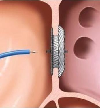
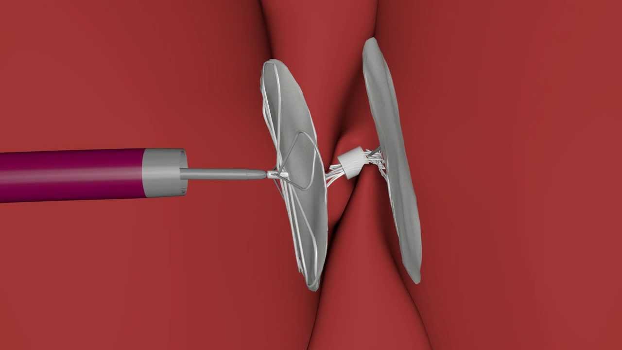
In the ASD closure process, a balloon is sent to this area. When the balloon sits on the hole, it is inflated and the diameter of the hole is measured, and then this balloon is removed. A closure device is delivered attached to the tip of a catheter. This device is in the form of two metal discs connected to each other in the middle. When the device is brought into the left atrium, first one of the discs is opened and rests on the part of the septum facing the left atrium, and the catheter is removed from the hole, and during this protrusion, the other disc of the device opens and leans against the wall facing the right atrium. Thus, the hole is closed. After imaging with transesophageal echocardiography, the catheter is removed from the vein.
After the Process;
After the procedure, the patient is taken to the room and a sand bag is placed on the operation area for 2 hours. After a 1-day stay in the hospital, the patient is discharged. After discharge, the first control is done in the 1st week. Subsequent checks are made in the 1st, 3rd, and 6th months. Uses aspirin for as long as deemed appropriate by the doctor. The top of the device that closes the hole is covered with endothelial tissue in a period of 6 months to 1 year. Afterwards, no medication is usually required.