
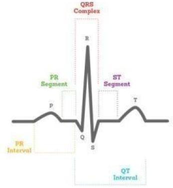
ECG, which is mostly applied to evaluate the risk of possible heart attack in people who apply to the doctor with chest pain complaints, is taken with the help of electrodes stuck on the skin. Thanks to the measurement of the electrical activity created by each contraction of the heart, detailed information about the heart is obtained. 10 electrodes are attached to the patient's body in order to measure the contraction and relaxation movement created by the atria and ventricles in the heart, the stimulation of the heart by cell groups, and the electrical activity that occurs during the transmission of this stimulus by ECG. These electrodes are connected to the EKG device with cables. The electrical activity data is converted into graphics by the EKG device and printed on a piece of paper called an electrocardiogram by the printer of the EKG device. This paper is 1x1 and 5x5 mm. consists of squares. 1x1mm. While graphs of electrical activity occurring every 0.04 seconds are printed on squares of size; On the other hand, the data of the activity occurring in 0.2 seconds are printed on the 5x5 mm squares. The EKG device records the heartbeats consisting of P, Q, R, S, T and U waves on the electrocardiogram. The graphical values created by these waves give the physician information about the heart health of the patient. The EKG device can be taken anywhere due to its easy portability. Thus, the inpatient does not need to be taken to the ECG room. The fact that it is a painless method and the duration of the procedure takes approximately 2-3 minutes provides convenience for both the patient and the physician. ECG, which enables rapid measurement of many data about heart health, is a diagnostic method frequently used by cardiologists and emergency physicians today.

How is an EKG taken?
Electrocardiography is mostly drawn to people who apply to a cardiologist with complaints such as chest pain, palpitations, shortness of breath, dizziness, blackouts and fainting. ECG, which is a completely safe test method without any risk, only records the electrical activity of the heart. Thanks to the ECG, which has become the gold standard in the evaluation of heart function, contraction of the heart muscle, rhythm and conduction disorders can be understood. The procedure, which does not require the patient to make any special preparations, is performed in a special room. Before the procedure, it is important to remove the jewelry on the neck and wrist. The patient is asked to remove the clothes above the waist or to pull them up if possible and lie on their back on the examination table. After cleaning the skin surface with a special solution for better adhesion of the electrodes, the electrodes are adhered to the chest, leg and arm areas. During 2-3 minutes of shooting, the patient is asked not to move or talk too much. After the shooting is completed, the data obtained by the EKG device while the person is being prepared is converted into a graphic and printed on a paper called an electrocardiogram. Pain and pain are not felt during the procedure, which does not require the use of any medication. The person can return to his daily life after the EKG, which does not cause any harm to the body. The physician evaluates the ECG result together with the examination findings. According to the data obtained, the physician may request additional examinations or arrange treatment. Electrocardiography also has different types such as stress EKG and Holter EKG.
What is a stress EKG?
Stress ECG, also known as heart stress test, exercise test or treadmill, records the electrical activity of the heart under load on the electrocardiogram instantly. Thanks to the effort test performed on the treadmill with a protocol, the reactions of the heart under pressure are measured. The exercise ECG, which is used in the investigation of cardiovascular diseases, is performed to determine whether the complaints of the patient who applied to the doctor with different complaints increase during exercise and to observe what kind of problem occurs in the heart in case of complaints. It is recommended to eat light foods before the exercise ECG procedure. Wearing comfortable and sports clothes also increases the comfort of the person during the test. Before the exercise test begins, the person is placed on the treadmill, where electrodes are attached to the skin surface. The exercise type is determined according to the age, gender and condition of the person to be tested for the exercise EKG. The test, which starts with brisk walking, includes exercises such as brisk walking, climbing hills, and running. With the exercise, it is aimed to increase the heart load. During all these exercises, heart movements are recorded by the EKG device. During exercise, blood pressure is also measured at certain periods. The data obtained during the exercise are sent to the specialist physician. The physician evaluates the exercise ECG test along with other examination findings. In the light of the information obtained, the physician may start treatment or request additional examinations.

What is Holter ECG?
Holter EKG, which is the size of a phone, is used in the presence of complaints such as palpitations, blackouts, fainting, and in the observation of heart rhythm disorders. This battery-operated device is attached to a person's waist or neck with a sling. The electrodes connected to the device are adhered to the chest area, on the skin. Unless there is a special situation, the person is asked to return to his normal daily life and do all the movements he normally does. At this point, the aim is to monitor the complaints that exist periodically. For 24 hours, the device measures the electrical activity created by the heart, as well as data such as pulse and blood pressure, and records these data. At the end of this period, the physician removes the holter ECG device and transfers the obtained data to the computer. According to the Holter ECG result, the physician observes what is happening in the heart during the current periodic complaints through graphics. Thanks to the Holter ECG, many discomforts that cannot be noticed during routine examination can be understood. Thus, the physician arranges the appropriate treatment.
Why is an EKG taken?
EKG, taken with the help of electrodes connected to the body, provides the physician with various information about the heart. Today, EKG is drawn to almost every person who applies to a cardiologist. The EKG procedure, which is applied to people who are admitted to the hospital with the suspicion of a heart attack, can also be taken in the following situations:
In the presence of suspicion arrhythmia or, in other words, rhythm and conduction disorder
In the presence of possible problems in the heart valves
In case of suspicion of narrowing or occlusion of the heart vessels defined as coronary arteries
In the presence of insufficient blood supply or damage to the heart, which is defined as ischemia
When the condition of the heart muscle, called hypertrophy, should be observed
Detection of imbalances in blood ions