
Coronary angioplasty is an attempt to open the diseased vessel that is narrowed or blocked. Coronary angioplasty, which is the process of opening a vessel with a balloon, is performed when blood flow is cut off due to narrowing or obstruction. It is a procedure that can be performed under local anesthesia. Thanks to this process, the obstacles in front of the blood flow that the heart needs are removed, and the blocked or narrowed part is expanded.
 In which cases is coronary angioplasty performed?
Coronary angioplasty is applied to patients with coronary artery disease, which develops due to the cessation of blood flow due to narrowing and blockages in the coronary arteries feeding the heart muscle. It is also applied in patients who have had a heart attack. Some complaints are seen due to the decrease in the amount of blood that occurs as a result of the narrowing of the vessels. These complaints can be as follows;
Tiredness
chest tightness
Combustion
Pain in left arm
Stomache ache
Pain radiating to the jaw
These complaints can be initiated by situations such as exercise and stress that increase the blood requirement of the heart, and usually the complaints go away on their own. Coronary angioplasty is preferred in cases where this method is appropriate in the narrowness of the patient's vascular structure and sufficient gain can be achieved, or in cases where the risk of surgical intervention is very high.
How is coronary angioplasty done?
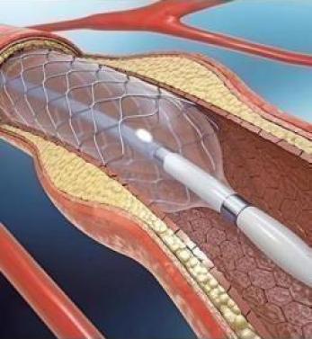
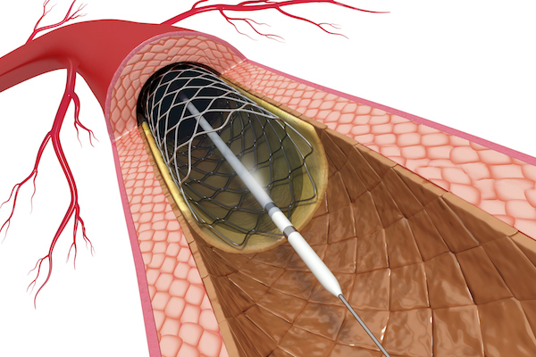
The thin balloon catheter is inflated by placing it in the narrowed area of the coronary artery. Thus, the plaque that causes the narrowing of the vessel is crushed and cracked open. Balloon angioplasty is similar to normal angioplasty. The patient is not placed under general anesthesia and the procedure is performed in the angio room. The patient's vein is entered through the right groin or wrist. Afterwards, a catheter is placed into the vein and dye is injected, allowing the vein to be seen. A thin wire is sent into the vein with the help of a catheter. If the vessel is completely occluded, the sent wire may not pass through the guide area. Afterwards, the balloon is sent over this wire and inflated in the narrow region of the vessel. Thus, the layers forming the stenosis are pushed to the vessel wall. Finally, the balloon is deflated and the vessel status is displayed.